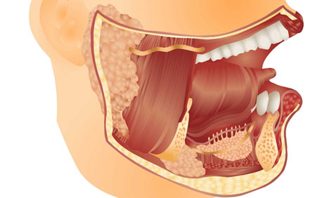
This is one of the salivary glands that is situated under your jaw. Approximately 60% of the lumps in the submandibular gland are benign (not cancerous) and the rest can be cancerous. They occur as a result of overgrowth of the cells in the gland. Swelling can also arise, as a result of stones blocking the duct draining the gland. This often leads to infection and pain, restricting routine activity.
Investigation and treatment of submandibular lumps
During the initial consultation a detailed history is taken and a thorough assessment is carried out. A needle may be inserted into the gland to collect a sample of cells from the lump. These cells are then analysed under the microscope by the pathologist to assess the nature of the swelling. If stones are suspected X-rays are often performed first. Occasionally, other tests such as a CT scan, MRI scan or a sialogram may be required.
Treatment depends on the nature of the lump and the results of the tests. Removal of the swelling is usually recommended because the exact nature of the swelling is often ascertained after removal and analysis of the whole lump. Additionally, if the lumps are not removed, the majority of them will grow further, often becoming cosmetically unacceptable and may even turn cancerous. Large, cancerous lumps are difficult to remove and complicate surgery.
Submandibular gland surgery explained
The operation involves removing the whole of the gland. This is performed under general anaesthetic, which means that you will be asleep throughout the procedure. An incision is made well below the jawline in the neck. The cut is usually placed along a skin crease so that over a period of time the scar is barely visible. At the end of the operation, a drain (plastic tube) is placed through the skin in order to prevent any blood or fluid collecting under the skin. This tube is usually removed the next day when you will be able to go home.
Potential complications
Weakness of the corner of the mouth
The nerve that moves the corner of the mouth lies in close proximity to the gland and is at risk during surgery. Damage to this nerve results in weakness of the corner of the mouth. This deformity is more obvious when one smiles. One may also experience drooling of saliva on the affected side. In most cases, the nerve works normally after surgery, although occasionally (about 10% of cases) you may notice a temporary weakness of the corner of the mouth. This usually lasts for a few weeks before full recovery takes place.
Numbness and stiffness of the neck
Stiffness and numbness of the neck are common and this resolves spontaneously over a period of few months. Use of moisturisers and creams to supple the scar and skin is very useful.
Blood and saliva collection
Blood and/or saliva can collect beneath the skin. Occasionally, it may be necessary to return to the operating theatre to remove this clot. Usually, this collection is minor and our body mops it away completely.
Altered taste
The nerve, which helps us appreciate taste, runs very close to the duct of the submandibular gland. This nerve may get bruised or damaged resulting in altered taste in the mouth. Usually, the altered taste sensation recovers fully over a period of few weeks.
Weakness of the tongue
Very rarely, the nerve that moves the tongue may get bruised/damaged during surgery. Usually this recovers fully over a period of a few weeks.


